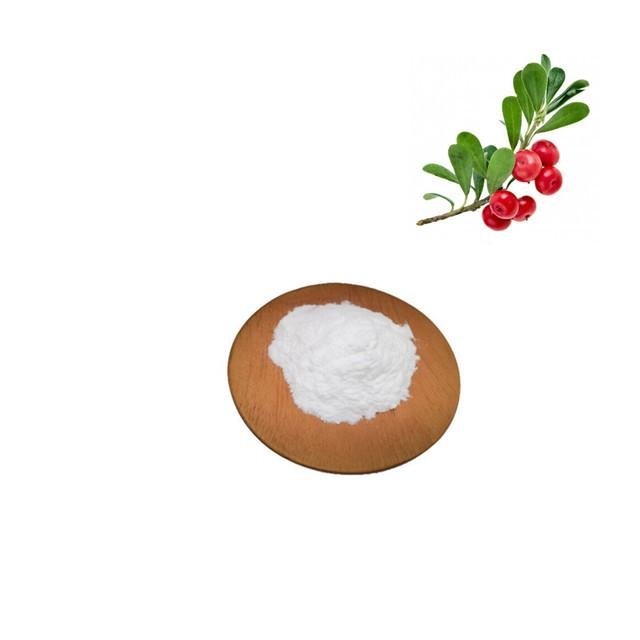
熊果甙
品名:Alpha arbutin
形式:粉末
Cas号.:84380-01-8
分子式:C12H16O7
分子量:272.25
外观:白色结晶粉末
检测方式:HPLC
规格:99%

阿尔法熊果苷,中文别名:4-氢苯醌-alpha-D-吡喃葡萄糖甙,对苯二酚-alpha-D-葡萄糖苷,或者对-羟基苯-α-D-吡喃葡萄糖苷,呈白色粉末或者白色结晶状。α-熊果苷是熊果苷的差相异构体,近年来发现它比熊果苷具有更强烈的酪氨酸酶抑制作用,能阻止黑色素的生成,从而减少皮肤色素沉积,祛除色斑和雀斑,而且对黑色素细胞不产生毒害性、刺激性、致敏性等副作用,同时还有杀菌、消炎的作用,从而引起了化妆品市场的广泛关注。α-熊果苷在很低的浓度下就能抑制酪氨酸酶的活性,虽然抑制机理不同于熊果苷,但其强度是熊果苷的近10倍,而且在较高的浓度下对细胞的生长也不产生影响。2002年以来欧美市场已经将α-熊果苷作为一种安全高效的亮肤活性剂推向高端化妆品市场,是二十一世纪最理想的皮肤美白祛斑活性剂。
主要用途: 用于高级化妆品中,可配制成护肤霜,高级珍珠霜等,既能美容护肤,又能消炎、抗刺激性。
烧烫伤药原料:熊果苷是新型烧烫伤药主要成分,特点是快速止痛,消炎力强,迅速消除红肿,愈合快,不留疤痕。
肠道消炎用药原料:杀菌、消炎效果好,无毒副作用。
产品性状 本品为白色结晶状。
产品规格 99.5%min
检测方法 HPLC
CAS 编码 84380-01-8
产品详询:13657416805
参考文献:
- 1.
Saeedi M, Eslamifar M, Khezri K. Kojic acid applications in cosmetic and pharmaceutical preparations. Biomedicine & Pharmacotherapy. 2019;110:582–93.
- 2.
Desmedt B, Courselle P, De Beer JO, Rogiers V, Grosber M, Deconinck E, et al. Overview of skin whitening agents with an insight into the illegal cosmetic market in Europe. J Eur Acad Dermatol Venereol. 2016;30(6):943–50.
- 3.
Couteau C, Coiffard L. Overview of skin whitening agents: drugs and cosmetic products. Cosmetics. 2016;3(3).
- 4.
Migas P, Krauze-Baranowska M. The significance of arbutin and its derivatives in therapy and cosmetics. Phytochem Lett. 2015;13:35–40.
- 5.
Sccs, Degen GH. Opinion of the Scientific Committee on Consumer safety (SCCS)—opinion on the safety of the use of α-arbutin in cosmetic products. Regul Toxicol Pharmacol. 2016;74:75–6.
- 6.
Won J, Park J. Improvement of arbutin trans-epidermal delivery using radiofrequency microporation. 2014.
- 7.
Alkilani AZ, McCrudden MT, Donnelly RF. Transdermal drug delivery: innovative pharmaceutical developments based on disruption of the barrier properties of the stratum corneum. Pharmaceutics. 2015;7(4):438–70.
- 8.
Mahato R. Chapter 13—microneedles in drug delivery. In: Mitra AK, Cholkar K, Mandal A, editors. Emerging nanotechnologies for diagnostics, drug delivery and medical devices. Boston: Elsevier; 2017. p. 331–53.
- 9.
Pamornpathomkul B, Ngawhirunpat T, Tekko IA, Vora L, McCarthy HO, Donnelly RF. Dissolving polymeric microneedle arrays for enhanced site-specific acyclovir delivery. Eur J Pharm Sci. 2018;121:200–9.
- 10.
Simon GA, Maibach HI. The pig as an experimental animal model of percutaneous permeation in man: qualitative and quantitative observations—an overview. Skin Pharmacol Appl Ski Physiol. 2000;13(5):229–34.
- 11.
Touitou E, Meidan VM, Horwitz E. Methods for quantitative determination of drug localized in the skin. J Control Release. 1998;56(1–3):7–21.
- 12.
Cilurzo F, Minghetti P, Sinico C. Newborn pig skin as model membrane in in vitro drug permeation studies: a technical note. AAPS PharmSciTech. 2007;8(4):E94-E.
- 13.
Davies DJ, Ward RJ, Heylings JR. Multi-species assessment of electrical resistance as a skin integrity marker for in vitro percutaneous absorption studies. Toxicol In Vitro. 2004;18(3):351–8.
- 14.
El-Say KM. Maximizing the encapsulation efficiency and the bioavailability of controlled-release cetirizine microspheres using Draper-Lin small composite design. Drug Des Dev Ther. 2016;10:825–39.
- 15.
Machekposhti SA, Soltani M, Najafizadeh P, Ebrahimi SA, Chen P. Biocompatible polymer microneedle for topical/dermal delivery of tranexamic acid. J Controlled Release. 2017;261:87–92.
- 16.
Larrañeta E, Moore J, Vicente-Pérez EM, Gonzalez Vazquez P, Lutton R, David Woolfson A, et al. A proposed model membrane and test method for microneedle insertion studies. 2014.
- 17.
Yao G, Quan G, Lin S, Peng T, Wang Q, Ran H, et al. Novel dissolving microneedles for enhanced transdermal delivery of levonorgestrel: in vitro and in vivo characterization. Int J Pharm. 2017;534(1–2):378–86.
- 18.
Structural characterization of inclusion complex of arbutin
- 19.
Larrañeta E, Henry M, Irwin NJ, Trotter J, Perminova AA, Donnelly RF. Synthesis and characterization of hyaluronic acid hydrogels crosslinked using a solvent-free process for potential biomedical applications. Carbohydr Polym. 2018;181:1194–205.
- 20.
LaFountaine JS, Prasad LK, Brough C, Miller DA, McGinity JW, Williams RO 3rd. Thermal processing of PVP- and HPMC-based amorphous solid dispersions. AAPS PharmSciTech. 2016;17(1):120–32.
- 21.
Baghel S, Cathcart H, O'Reilly NJ. Polymeric amorphous solid dispersions: a review of amorphization, crystallization, stabilization, solid-state characterization, and aqueous solubilization of biopharmaceutical classification system class II drugs. J Pharm Sci. 2016;105(9):2527–44.
- 22.
Zhang M, Ma Y, Wang Z, Han Z, Gao W, Gu Y. Optimizing molecular weight of octyl chitosan as drug carrier for improving tumor therapeutic efficacy. Oncotarget. 2017;8(38):64237–49.
- 23.
Kariduraganavar MY, Kittur AA, Kamble RR. Chapter 1—polymer synthesis and processing. In: Kumbar SG, Laurencin CT, Deng M, editors. Natural and synthetic biomedical polymers. Oxford: Elsevier; 2014. p. 1–31.
- 24.
Tritt-Goc J, Kowalczuk J, Pislewski N. Hydration of hydroxypropylmethyl cellulose: effects of pH and molecular mass. Acta Physica Polonica A - Acta Phys Pol A. 2006;108.
- 25.
Sarkar N, Walker LC. Hydration—dehydration properties of methylcellulose and hydroxypropylmethylcellulose. Carbohydr Polym. 1995;27(3):177–85.
- 26.
Chen CP, Hsieh CM, Tsai T, Yang JC, Chen CT. Optimization and evaluation of a chitosan/hydroxypropyl methylcellulose hydrogel containing toluidine blue O for antimicrobial photodynamic inactivation. Int J Mol Sci. 2015;16(9):20859–72.
- 27.
Mojumdar EH, Pham QD, Topgaard D, Sparr E. Skin hydration: interplay between molecular dynamics, structure and water uptake in the stratum corneum. Sci Rep. 2017;7(1):15712.
- 28.
Verdier-Sevrain S, Bonte F. Skin hydration: a review on its molecular mechanisms. J Cosmet Dermatol. 2007;6(2):75–82.
- 29.
Shabbir M, Ali S, Raza M, Sharif A, Akhtar MF, Manan A, et al. Effect of hydrophilic and hydrophobic polymer on in vitro dissolution and permeation of bisoprolol fumarate through transdermal patch. Acta Pol Pharm. 2017;74:187–97.
- 30.
Grubauer G, Elias PM, Feingold KR. Transepidermal water loss: the signal for recovery of barrier structure and function. J Lipid Res. 1989;30(3):323–33.
- 31.
Menon GK, Feingold KR, Elias PM. Lamellar body secretory response to barrier disruption. J Investig Dermatol. 1992;98(3):279–89
- 32.
Gupta J, Gill HS, Andrews SN, Prausnitz MR. Kinetics of skin resealing after insertion of microneedles in human subjects. J Control Release. 2011;154(2):148–55.