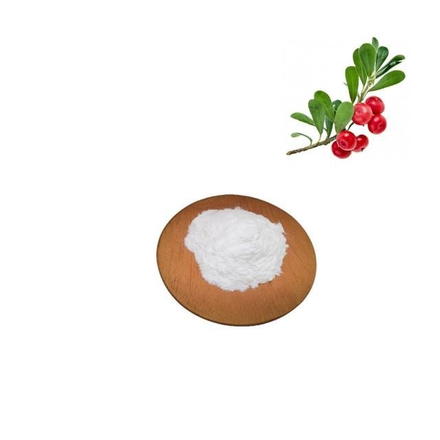
Alpha arbutin for cosmetics
Product name:Alpha arbutin
Form:Powder
Cas No.:84380-01-8
Molecular Formula:C12H16O7
Molecular Weight:272.25
Appearance:White crystalline powder
Test Method:HPLC
Specification:99%

About Aplha Arbutin
Alpha Arbutin is the active substance originated from natural plant which can whiten and lighten skin. Alpha Arbutin Powder can infiltrate into the skin quickly without affecting the concentration of cell multiplication and effectively prevent activity of tyrosinase in the skin and the forming of melanin. By combined arbutin with tyrosinase, decomposition and drainage of melanin are accelerated, splash and fleck can be got ride of and no side effects are caused.Arbutin Powder is one of the safest and most efficient whitening materials that are popular at present. Alpha Arbutin is also the most competitive whitening activity in the 21st century.
Alpha-arbutin is similar to β-arbutin, which inhibits the production and deposition of melanin and eliminates spots and freckles. Studies have shown that α-arbutin can inhibit the activity of tyrosinase at a lower concentration, and its inhibition of tyrosinase is better than that of β arbutin.
Alpha arbutin main function:
(1) Alpha arbutin powder can protect the skin against damage caused by free radicals.have the function of anti-aging
(2) Alpha arbutin powder is a skin whitening agent
(3) Alpha arbutin powder inhibits the formation of melanin pigment by inhibiting Tyrosinase activity.
2.Medical Field Function
(1) Alpha arbutin powder was first used in medical areas as an anti-inflammatory and antibacterial agent.
(2) Alpha arbutin powderwas used particularly for cystitis, urethritis and pyelitis.
(3) Alpha arbutin powder may be used to repress the virulence of bacterial pathogens and to prevent contaminating bacteria.
(4) Alpha arbutin powder is used for treating allergic inflammation of the skin .
Alpha arbutin application:
1.Alpha arbutin can be used in cosmetic field.
1.Alpha arbutin can be used in medical field.
For more product information pls kindly contact email sales09@staherb.cn
Alpha arbutin product analysis
| Product Name |
Alpha Arbutin/Bearberry Extract |
Appearance |
White Crystal Powder |
||
| Cas No. |
84380-01-8 |
Odor |
Characteristic |
||
| Molecular Formula |
C12H16O7 |
Molecular Weight |
272.25 |
||
| Standard |
USP/BP |
||||
| Shelf Life |
Two Years |
||||
| Storage |
Store in sealed containers at cool & dry place. Protect from sunlight, moisture and pest infestation. |
||||
| Item |
Specification |
Results |
| Appearance |
White crystal powder |
White Crystal Powder |
| Assay |
99.0%Min |
99.9% |
| Melting point |
203~206±1°C |
205-206 |
| Clarity of water solution |
Transparency, colorless, none suspended matters |
Pass |
| pH value of 1% aqueous solution |
5.0~7.0 |
6.90 |
| Specific optical rotation |
[α]D20=+176~184º |
+178.8 |
| Arsenic |
≤2 ppm |
pass |
| Hydroquinone |
≤10 ppm |
pass |
| Heavy metal |
≤10 ppm |
pass |
| Loss on drying |
≤0.5% |
0.07% |
| Ignition residue |
≤0.5% |
pass |
| Pathogen |
Bacteria:≤100cfu/g Fungus:≤100cfu/g |
Pass |
References:
- 1.
Saeedi M, Eslamifar M, Khezri K. Kojic acid applications in cosmetic and pharmaceutical preparations. Biomedicine & Pharmacotherapy. 2019;110:582–93.
- 2.
Desmedt B, Courselle P, De Beer JO, Rogiers V, Grosber M, Deconinck E, et al. Overview of skin whitening agents with an insight into the illegal cosmetic market in Europe. J Eur Acad Dermatol Venereol. 2016;30(6):943–50.
- 3.
Couteau C, Coiffard L. Overview of skin whitening agents: drugs and cosmetic products. Cosmetics. 2016;3(3).
- 4.
Migas P, Krauze-Baranowska M. The significance of arbutin and its derivatives in therapy and cosmetics. Phytochem Lett. 2015;13:35–40.
- 5.
Sccs, Degen GH. Opinion of the Scientific Committee on Consumer safety (SCCS)—opinion on the safety of the use of α-arbutin in cosmetic products. Regul Toxicol Pharmacol. 2016;74:75–6.
- 6.
Won J, Park J. Improvement of arbutin trans-epidermal delivery using radiofrequency microporation. 2014.
- 7.
Alkilani AZ, McCrudden MT, Donnelly RF. Transdermal drug delivery: innovative pharmaceutical developments based on disruption of the barrier properties of the stratum corneum. Pharmaceutics. 2015;7(4):438–70.
- 8.
Mahato R. Chapter 13—microneedles in drug delivery. In: Mitra AK, Cholkar K, Mandal A, editors. Emerging nanotechnologies for diagnostics, drug delivery and medical devices. Boston: Elsevier; 2017. p. 331–53.
- 9.
Pamornpathomkul B, Ngawhirunpat T, Tekko IA, Vora L, McCarthy HO, Donnelly RF. Dissolving polymeric microneedle arrays for enhanced site-specific acyclovir delivery. Eur J Pharm Sci. 2018;121:200–9.
- 10.
Simon GA, Maibach HI. The pig as an experimental animal model of percutaneous permeation in man: qualitative and quantitative observations—an overview. Skin Pharmacol Appl Ski Physiol. 2000;13(5):229–34.
- 11.
Touitou E, Meidan VM, Horwitz E. Methods for quantitative determination of drug localized in the skin. J Control Release. 1998;56(1–3):7–21.
- 12.
Cilurzo F, Minghetti P, Sinico C. Newborn pig skin as model membrane in in vitro drug permeation studies: a technical note. AAPS PharmSciTech. 2007;8(4):E94-E.
- 13.
Davies DJ, Ward RJ, Heylings JR. Multi-species assessment of electrical resistance as a skin integrity marker for in vitro percutaneous absorption studies. Toxicol In Vitro. 2004;18(3):351–8.
- 14.
El-Say KM. Maximizing the encapsulation efficiency and the bioavailability of controlled-release cetirizine microspheres using Draper-Lin small composite design. Drug Des Dev Ther. 2016;10:825–39.
- 15.
Machekposhti SA, Soltani M, Najafizadeh P, Ebrahimi SA, Chen P. Biocompatible polymer microneedle for topical/dermal delivery of tranexamic acid. J Controlled Release. 2017;261:87–92.
- 16.
Larrañeta E, Moore J, Vicente-Pérez EM, Gonzalez Vazquez P, Lutton R, David Woolfson A, et al. A proposed model membrane and test method for microneedle insertion studies. 2014.
- 17.
Yao G, Quan G, Lin S, Peng T, Wang Q, Ran H, et al. Novel dissolving microneedles for enhanced transdermal delivery of levonorgestrel: in vitro and in vivo characterization. Int J Pharm. 2017;534(1–2):378–86.
- 18.
Structural characterization of inclusion complex of arbutin
- 19.
Larrañeta E, Henry M, Irwin NJ, Trotter J, Perminova AA, Donnelly RF. Synthesis and characterization of hyaluronic acid hydrogels crosslinked using a solvent-free process for potential biomedical applications. Carbohydr Polym. 2018;181:1194–205.
- 20.
LaFountaine JS, Prasad LK, Brough C, Miller DA, McGinity JW, Williams RO 3rd. Thermal processing of PVP- and HPMC-based amorphous solid dispersions. AAPS PharmSciTech. 2016;17(1):120–32.
- 21.
Baghel S, Cathcart H, O'Reilly NJ. Polymeric amorphous solid dispersions: a review of amorphization, crystallization, stabilization, solid-state characterization, and aqueous solubilization of biopharmaceutical classification system class II drugs. J Pharm Sci. 2016;105(9):2527–44.
- 22.
Zhang M, Ma Y, Wang Z, Han Z, Gao W, Gu Y. Optimizing molecular weight of octyl chitosan as drug carrier for improving tumor therapeutic efficacy. Oncotarget. 2017;8(38):64237–49.
- 23.
Kariduraganavar MY, Kittur AA, Kamble RR. Chapter 1—polymer synthesis and processing. In: Kumbar SG, Laurencin CT, Deng M, editors. Natural and synthetic biomedical polymers. Oxford: Elsevier; 2014. p. 1–31.
- 24.
Tritt-Goc J, Kowalczuk J, Pislewski N. Hydration of hydroxypropylmethyl cellulose: effects of pH and molecular mass. Acta Physica Polonica A - Acta Phys Pol A. 2006;108.
- 25.
Sarkar N, Walker LC. Hydration—dehydration properties of methylcellulose and hydroxypropylmethylcellulose. Carbohydr Polym. 1995;27(3):177–85.
- 26.
Chen CP, Hsieh CM, Tsai T, Yang JC, Chen CT. Optimization and evaluation of a chitosan/hydroxypropyl methylcellulose hydrogel containing toluidine blue O for antimicrobial photodynamic inactivation. Int J Mol Sci. 2015;16(9):20859–72.
- 27.
Mojumdar EH, Pham QD, Topgaard D, Sparr E. Skin hydration: interplay between molecular dynamics, structure and water uptake in the stratum corneum. Sci Rep. 2017;7(1):15712.
- 28.
Verdier-Sevrain S, Bonte F. Skin hydration: a review on its molecular mechanisms. J Cosmet Dermatol. 2007;6(2):75–82.
- 29.
Shabbir M, Ali S, Raza M, Sharif A, Akhtar MF, Manan A, et al. Effect of hydrophilic and hydrophobic polymer on in vitro dissolution and permeation of bisoprolol fumarate through transdermal patch. Acta Pol Pharm. 2017;74:187–97.
- 30.
Grubauer G, Elias PM, Feingold KR. Transepidermal water loss: the signal for recovery of barrier structure and function. J Lipid Res. 1989;30(3):323–33.
- 31.
Menon GK, Feingold KR, Elias PM. Lamellar body secretory response to barrier disruption. J Investig Dermatol. 1992;98(3):279–89
- 32.
Gupta J, Gill HS, Andrews SN, Prausnitz MR. Kinetics of skin resealing after insertion of microneedles in human subjects. J Control Release. 2011;154(2):148–55.