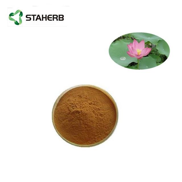
lotus leaf extract Nuciferine
Product Name:Lotus Leaf Extract
Latin Name:Folium Nelumbinis
Active Ingredient:Nuciferine 1% 2% 5% 10% 50%,98%
Test Method:HPLC
Extraction Solvent:Ethanol/ Water
CAS NO:475-83-2
Molecular Formula:C19H21NO2
Molecular Weight:313.39082

About lotus leaf
Lotus leaf is a flowering aquatic perennial that grows widely throughout tropical regions of Asia and the Middle East. The pale green leaves are flat and broad, reaching up to 18 inches in diameter. The leaves are usually collected in the summer and autumn and dried before being cut into small pieces, powdered or processed into pills. Lotus leaves are touted to be useful in treating a variety of conditions, including heavy bleeding, diarrhea and muscle spasms.
Louts leaf extract Function:
1. Lotus leaf extract has the function of weight control.
2. Lotus leaf extract can adjust blood lipids, and expectorant.
3. Lotus leaf extract is used as anticoagulant and antidote in medicine.
4. Lotus leaf extract has strong effect on lowering the blood pressure
5. Lotus leaf P.E. has become popular to lower blood cholesterol
6. Lotus leaf P.E. can treat fatty liver and promote blood circulation.
Lotus leaf extract Application:
1. Applied in food field, lotus leaf become a delicious and healthy green food;
2. Applied in health product field, lotus leaf slimming tea by the majority of ladys love;
3. Applied in pharmaceutical field, it can promote the growth of bone.
4. Applied in Dietary Supplement, it is greatest fat burner, reduce fat, depress blood pressure.
For more product information pls contact email sales09@staherb.cn
Specification:
| Item |
Specification |
Method Result |
|
| Physical Property |
|
|
|
| Appearance |
Fine Powder |
Organoleptic Conforms |
|
| Color |
Brown powder |
Organoleptic Conforms |
|
| Odour |
Characteristic |
Organoleptic Conforms |
|
| General Analysis |
|
|
|
| Identification Ratio Loss on Drying |
Identical to R.S. sample 10:1 <5.0%c |
TLC TLC Eur.Ph.6.0[2.8.17] |
Conforms Conforms 3.22% |
| Ash |
≤5.0% |
Eur.Ph.6.0[2.4.16] 3.37% |
|
| Contaminants |
|
|
|
| Pesticides Residue |
Meet USP32<561> |
USP32<561> Conforms |
|
| Lead(Pb) |
≤3.0mg/kg |
Eur.Ph6.0<2.2.58>ICP-MS 2.1 |
|
| Arsenic(As) |
≤2.0mg/kg |
Eur.Ph6.0<2.2.58>ICP-MS 1.9 |
|
| Cadmium(Cd) |
≤1.0mg/kg |
Eur.Ph6.0<2.2.58>ICP-MS 0.7 |
|
| Mercury(Hg) |
≤0.1mg/kg |
Eur.Ph6.0<2.2.58>ICP-MS 0.07 |
|
| Microbiological |
|
|
|
| Total Plate Count |
≤1000cfu/g |
USP30<61> 119 |
|
| Yeast &Mold |
≤100cfu/g |
USP30<61> 41 |
|
| E.Coli. |
Negative |
USP30<62> Conforms |
|
| Salmonella |
Negative |
USP30<62> Conforms |
|
References:
- 1.
Zhang X, Xu R, Zhang C, Xu Y, Han M, Huang B, et al. Trifluoperazine, a novel autophagy inhibitor, increases radiosensitivity in glioblastoma by impairing homologous recombination. J Exp Clin Cancer Res. 2017;36(1):118.
- 2.
Vredenburgh JJ, Desjardins A, Herndon JE 2nd, Dowell JM, Reardon DA, Quinn JA, et al. Phase II trial of bevacizumab and irinotecan in recurrent malignant glioma. Clin Cancer Res. 2007;13(4):1253–9.
- 3.
Kim BM, Hong Y, Lee S, Liu P, Lim JH, Lee YH, et al. Therapeutic implications for overcoming radiation resistance in cancer therapy. Int J Mol Sci. 2015;16(11):26880–913.
- 4.
Brabletz T, Kalluri R, Nieto MA, Weinberg RA. EMT in cancer. Nat Rev Cancer. 2018;18(2):128–34.
- 5.
Nieto MA, Huang RY, Jackson RA, Thiery JP. EMT: 2016. Cell. 2016;166(1):21–45.
- 6.
Shibue T, Weinberg RA. EMT, CSCs, and drug resistance: the mechanistic link and clinical implications. Nat Rev Clin Oncol. 2017 Oct;14(10):611–29.
- 7.
Phillips S, Kuperwasser C. SLUG: critical regulator of epithelial cell identity in breast development and cancer. Cell Adhes Migr. 2014;8(6):578–87.
- 8.
Voutsadakis IA. Epithelial-mesenchymal transition (EMT) and regulation of EMT factors by steroid nuclear receptors in breast cancer: a review and in silico investigation. J Clin Med. 2016;5(1):E11.
- 9.
Alves CC, Carneiro F, Hoefler H, Becker KF. Role of the epithelial-mesenchymal trasition regulator Slug in primary human cancers. Front Biosci (Landmark Ed). 2009;14:3035–50.
- 10.
Wegner M. From head to toes: the multiple facets of sox proteins. Nucleic Acids Res. 1999;27(6):1409–20.
- 11.
Mansouri S, Nejad R, Karabork M, Ekinci C, Solaroglu I, Aldape KD, et al. Sox2: regulation of expression and contribution to brain tumors. CNS Oncol. 2016;5(3):159–73.
- 12.
Song WS, Yang YP, Huang CS, Lu KH, Liu WH, Wu WW, et al. Sox2, stemness gene, regulates tumor-initiating and drug-resistant properties in CD133-positive glioblastoma stem cells. J Chin Med Assoc. 2016;79(10):538–45.
- 13.
Garros-Regulez L, Garcia I, Carrasco-Garcia E, Lantero A, Aldaz P, Moreno-Cugnon L, et al. Targeting SOX2 as a therapeutic strategy in glioblastoma. Front Oncol. 2016;6:222.
- 14.
Chin YW, Yoon KD, Kim J. Cytotoxic anticancer candidates from terrestrial plants. Anti Cancer Agents Med Chem. 2009;9(8):913–42.
- 15.
Mangal M, Sagar P, Singh H, Raghava GP, Agarwal SM. NPACT: naturally occurring plant-based anti-cancer compound-activity-target database. Nucleic Acids Res. 2013;41:D1124–9.
- 16.
Nguyen KH, Ta TN, Pham TH, Nguyen QT, Pham HD, Mishra S, et al. Nuciferine stimulates insulin secretion from beta cells-an in vitro comparison with glibenclamide. J Ethnopharmacol. 2012;142(2):488–95.
- 17.
Ho HH, Hsu LS, Chan KC, Chen HM, Wu CH, Wang CJ. Extract from the leaf of nucifera reduced the development of atherosclerosis via inhibition of vascular smooth muscle cell proliferation and migration. Food Chem Toxicol. 2010;48(1):159–68.
- 18.
Kashiwada Y, Aoshima A, Ikeshiro Y, Chen YP, Furukawa H, Itoigawa M, et al. Anti-HIV benzylisoquinoline alkaloids and flavonoids from the leaves of Nelumbo nucifera, and structure-activity correlations with related alkaloids. Bioorg Med Chem. 2005;13(2):443–8.
- 19.
Nakamura S, Nakashima S, Tanabe G, Oda Y, Yokota N, Fujimoto K, et al. Alkaloid constituents from flower buds and leaves of sacred lotus (Nelumbo nucifera, Nymphaeaceae) with melanogenesis inhibitory activity in B16 melanoma cells. Bioorg Med Chem. 2013;21(3):779–87.
- 20.
Liu W, Yi DD, Guo JL, Xiang ZX, Deng LF, He L. Nuciferine, extracted from Nelumbo nucifera Gaertn, inhibits tumor-promoting effect of nicotine involving Wnt/β-catenin signaling in non-small cell lung cancer. J Ethnopharmacol. 2015;165:83–93.
- 21.
Guo F, Yang X, Li X, Feng R, Guan C, Wang Y, et al. Nuciferine prevents hepatic steatosis and injury induced by a high-fat diet in hamsters. PLoS One. 2013;8(5):e63770.
- 22.
Qi Q, Li R, Li HY, Cao YB, Bai M, Fan XJ, et al. Identification of the anti-tumor activity and mechanisms of nuciferine through a network pharmacology approach. Acta Pharmacol Sin. 2016;37(7):963–72.
- 23.
Xu Y, Bao S, Tian W, Wen C, Hu L, Lin C. Tissue distribution model and pharmacokinetics of nuciferine based on UPLC-MS/MS and BP-ANN. Int J Clin Exp Med. 2015;8(10):17612–22.
- 24.
Kastan MB, Bartek J. Cell-cycle checkpoints and cancer. Nature. 2004;432(7015):316–23.
- 25.
Sherr CJ. Cancer cell cycles. Science. 1996;274(5293):1672–7.
- 26.
Wang Y, Decker SJ, Sebolt-Leopold J. Knockdown of Chk1, Wee1 and Myt1 by RNA interference abrogates G2 checkpoint and induces apoptosis. Cancer Biol Ther. 2004;3(3):305–13.
- 27.
Fletcher L, Cheng Y, Muschel RJ. Abolishment of the Tyr-15 inhibitory phosphorylation site on cdc2 reduces the radiation-induced G(2) delay, revealing a potential checkpoint in early mitosis. Cancer Res. 2002;62(1):241–50.
- 28.
Junyan P, Shujuan Y, Shulin G, Yan C, Xia X. The antitumor effect of DYC-279 on human hepatocellular carcinoma HepG2 cells. Pharmacology. 2016;97(3–4):177–83.
- 29.
Stark GR, Taylor WR. Control of the G2/M transition. Mol Biotechnol. 2006;32(3):227–48.
- 30.
Glotzer M, Murray AW, Kirschner MW. Cyclin is degraded by the ubiquitin pathway. Nature. 1991;349(6305):132–8.
- 31.
Hershko A. Roles of ubiquitin-mediated proteolysis in cell cycle control. Curr Opin Cell Biol. 1997;9(6):788–99.
- 32.
Parry DH, O'Farrell PH. The schedule of destruction of three mitotic cyclins can dictate the timing of events during exit from mitosis. Curr Biol. 2001;11(9):671–83.
- 33.
Vorlaufer E, Peters JM. Regulation of the cyclin B degradation system by an inhibitor of mitotic proteolysis. Mol Biol Cell. 1998;9(7):1817–31.
- 34.
Lin H, Liu XY, Subramanian B, Nakeff A, Valeriote F, Chen BD. Mitotic arrest induced by XK469, a novel antitumor agent, is correlated with the inhibition of cyclin B1 ubiquitination. Int J Cancer. 2002;97(1):121–8.
- 35.
Lee YM, Lim DY, Choi HJ, Jung JI, Chung WY, Park JH. Induction of cell cycle arrest in prostate cancer cells by the dietary compound isoliquiritigenin. J Med Food. 2009;12(1):8–14.
- 36.
Chien CC, Wu MS, Shen SC, Ko CH, Chen CH, Yang LL, et al. Activation of JNK contributes to evodiamine-induced apoptosis and G2/M arrest in human colorectal carcinoma cells: a structure-activity study of evodiamine. PLoS One. 2014;9(6):e99729.
- 37.
Yang L, Liang H, Wang Y, Gao S, Yin K, Liu Z, et al. MiRNA-203 suppresses tumor cell proliferation, migration and invasion by targeting Slug in gastric cancer. Protein Cell. 2016;7(5):383–7.
- 38.
Vitali R, Mancini C, Cesi V, Tanno B, Mancuso M, Bossi G, et al. Slug (SNAI2) down-regulation by RNA interference facilitates apoptosis and inhibits invasive growth in neuroblastoma preclinical models. Clin Cancer Res. 2008;14(14):4622–30.
- 39.
Zhao X, Sun B, Sun D, Liu T, Che N, Gu Q, et al. Slug promotes hepatocellular cancer cell progression by increasing SOX2 and NANOG expression. Oncol Rep. 2015;33(1):149–56.
- 40.
Yang CY, Chen YD, Guo W, Gao Y, Song CQ, Zhang Q, et al. Bismuth ferrite-based nanoplatform design: an ablation mechanism study of solid tumor and NIR-triggered photothermal/photodynamic combination cancer therapy. Adv Funct Mater. 2018;28:1706827.
- 41.
Ke D, Yang R, Jing L. Combined diagnosis of breast cancer in the early stage by MRI and detection of gene expression. Exp Ther Med. 2018;16(2):467–72.
- 42.
Yang HW, Menon LG, Black PM, Carroll RS, Johnson MD. SNAI2/Slug promotes growth and invasion in human gliomas. BMC Cancer. 2010;10:301.
- 43.
Carpenter RL, Paw I, Dewhirst MW, Lo HW. Akt phosphorylates and activates HSF-1 independent of heat shock, leading to Slug overexpression and epithelial-mesenchymal transition (EMT) of HER2-overexpressing breast cancer cells. Oncogene. 2015;34(5):546–57.
- 44.
Yao C, Su L, Shan J, Zhu C, Liu L, Liu C, et al. IGF/STAT3/NANOG/Slug signaling axis simultaneously controls epithelial-mesenchymal transition and stemness maintenance in colorectal cancer. Stem Cells. 2016;34(4):820–31.
- 45.
Chen YD, Zhang Y, Dong TX, Xu YT, Zhang W, An TT, et al. Hyperthermia with different temperatures inhibits proliferation and promotes apoptosis through the EGFR/STAT3 pathway in C6 rat glioma cells. Mol Med Rep. 2017;16(6):9401–8.
- 46.
Brennan CW, Verhaak RG, McKenna A, Campos B, Noushmehr H, Salama SR, et al. The somatic genomic landscape of glioblastoma. Cell. 2013;155(2):462–77.
- 47.
Safa AR, Saadatzadeh MR, Cohen-Gadol AA, Pollok KE, Bijangi-Vishehsaraei K. Emerging targets for glioblastoma stem cell therapy. J Biomed Res. 2016;30(1):19–31.
- 48.
van Schaijik B, Davis PF, Wickremesekera AC, Tan ST, Itinteang T. Subcellular localisation of the stem cell markers OCT4, SOX2, NANOG, KLF4 and c-MYC in cancer: a review. J Clin Pathol. 2018;71(1):88–91.
- 49.
Takahashi K, Yamanaka S. Induction of pluripotent stem cells from mouse embryonic and adult fibroblast cultures by defined factors. Cell. 2006;126(4):663–76.
- 50.
Azuaje F, Tiemann K, Niclou SP. Therapeutic control and resistance of the EGFR-driven signaling network in glioblastoma. Cell Commun Signal. 2015;13:23.
- 51.
Liu Y, Wu X, Mi Y, Zhang B, Gu S, Liu G, et al. PLGA nanoparticles for the oral delivery of nuciferine: preparation, physicochemical characterization and in vitro/in vivo studies. Drug Deliv. 2017;24(1):443–51.